Figure 6 from Femoral Hernia: A Review of the Clinical Anatomy and

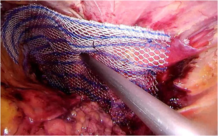
Figure 6. Femoral hernia repair in clean operation. (a) The narrow side of the mesh is sutured to Cooper’s ligament; (b) The mesh is sutured to the iliopubic tract or shelving portion of the inguinal ligament; (c) The posterior wall of the inguinal canal is reinforced, as in Lichtenstein’s repair. - "Femoral Hernia: A Review of the Clinical Anatomy and Surgical Treatment"
Anatomy essentials for laparoscopic inguinal hernia repair - Yang - Annals of Translational Medicine

PDF] Laparoscopic repair of an incarcerated femoral hernia
Frontiers Publishing Partnerships Primary Lumbar Hernia, Review and Proposals for a Standardized Treatment
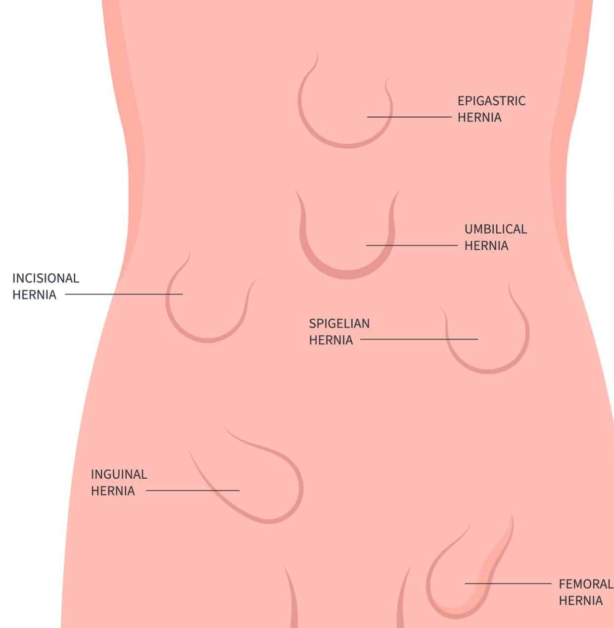
Abdominal Hernia - Epigastric - Spigelian - Obturator - TeachMeSurgery

PDF) Femoral Hernia: A Review of the Clinical Anatomy and Surgical Treatment
Clinical Anatomy of the Groin: Posterior Laparoscopic Approach

An intraoperative image showing an erythematous appendix found within
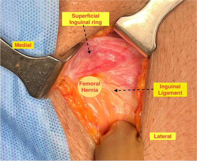
Left side femoral hernia – view from inside the abdomen (arrow)

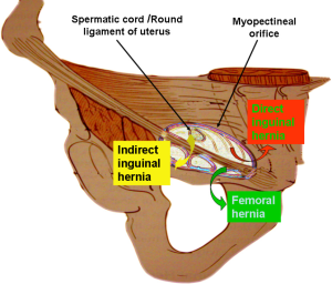
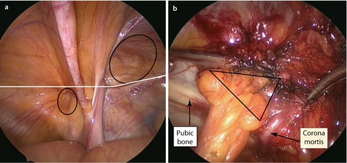
Anterior and posterior views of myopectineal orifice ( from Elliott and

Figure 7 from Femoral Hernia: A Review of the Clinical Anatomy and Surgical Treatment
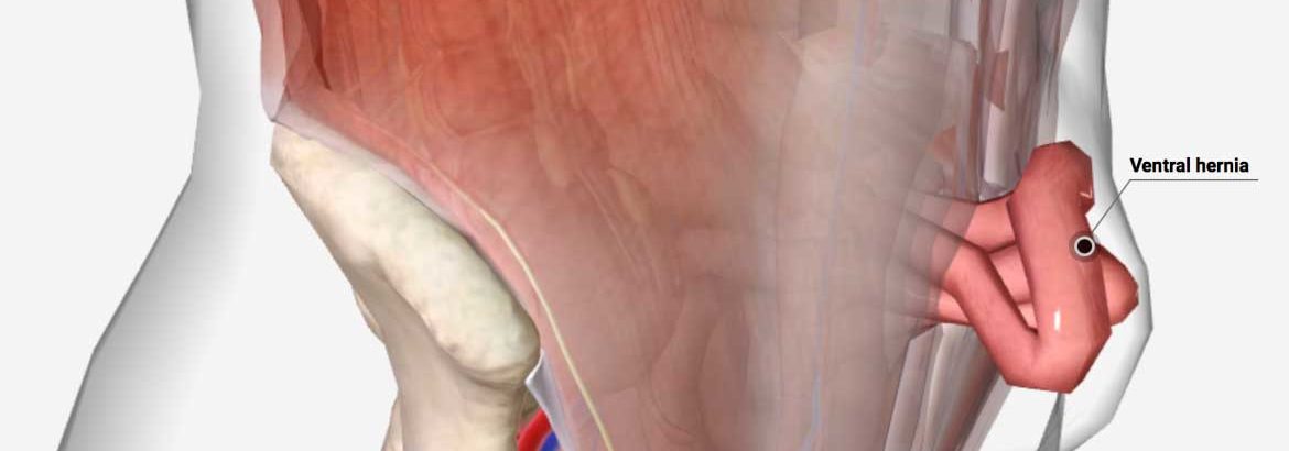
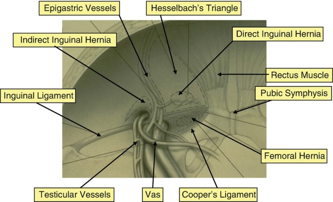
Femoral hernia anatomy

AIS Channel · Hernia Surgery