Beware of reflectance confocal microscopy artifacts when searching

Semantic segmentation of reflectance confocal microscopy mosaics of pigmented lesions using weak labels

Anais Brasileiros de Dermatologia, Vol 95, Issue 1, Pages 1-132 (January–February 2020)

White piedra, black piedra, tinea versicolor, and tinea nigra: contribution to the diagnosis of superficial mycosis. - Abstract - Europe PMC

Jean PERROT, Medical Doctor, Professor, Centre Hospitalier Universitaire de Saint-Étienne, Saint-Étienne, CHU St Etienne, Department of Dermatology

Jean PERROT, Medical Doctor, Professor, Centre Hospitalier Universitaire de Saint-Étienne, Saint-Étienne, CHU St Etienne, Department of Dermatology
The challenge of Diagnosing Common Dermatomycosis by Reflectance Confocal Microscopy

Learn To Minimize Artifacts In Fluorescence Microscopy

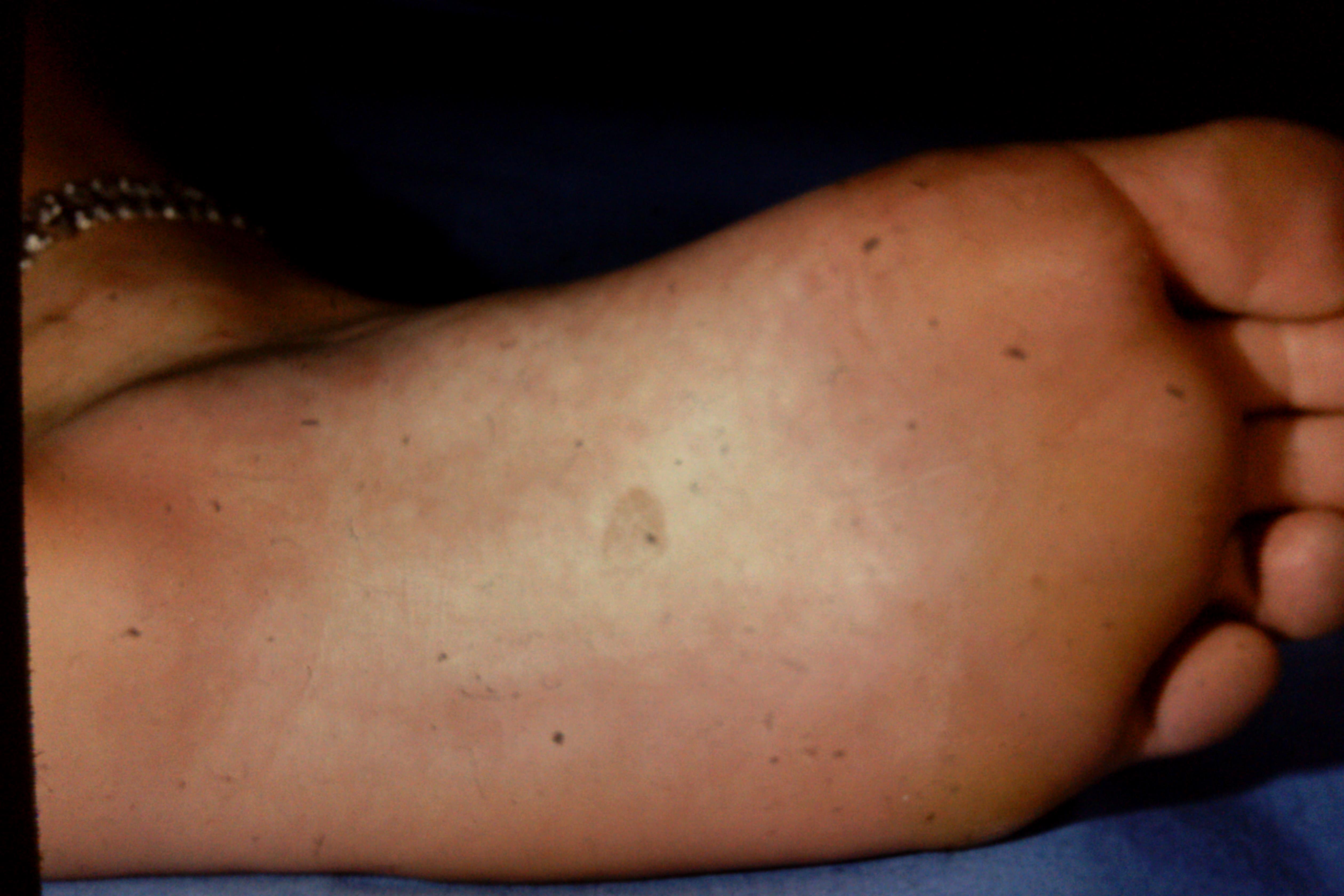
Two cases of tinea nigra with classic clinical presentation (A1, B1).

Elisa CINOTTI, Medical Doctor, Dermatologist, Azienda Ospedaliera Universitaria Senese, Siena, Dermatology

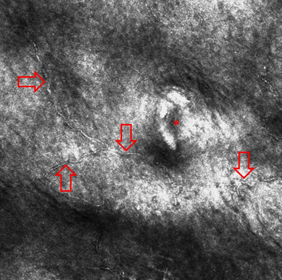
Grain density (~o) over fungal structures in relation to the grain